
Freeze, image, cure
Penn researchers are using electron microscopes to decode disease biology.
Nicholas Palmer was investigating a biological mystery: How does actin, a common protein that plays a role in the spread of cancer in the body, lengthen and shorten during cellular movement?
Individual proteins are too small to see with most microscopes, and other techniques had provided inconsistent clues to the precise mechanism.
So Palmer, a graduate student at the Perelman School of Medicine at the University of Pennsylvania, turned to a relatively new and extremely powerful electron microscope that provides some of the sharpest-ever images of life’s tiniest components.
He loaded millions of copies of frozen proteins into the sample chamber of the microscope, a machine the size of an industrial refrigerator, then waited as the images developed on the computer screen.
“When I saw this structure in particular, I jumped up and cheered,” Palmer said. “As soon as I saw this, I was like, ‘I know exactly what the mechanism is.’”
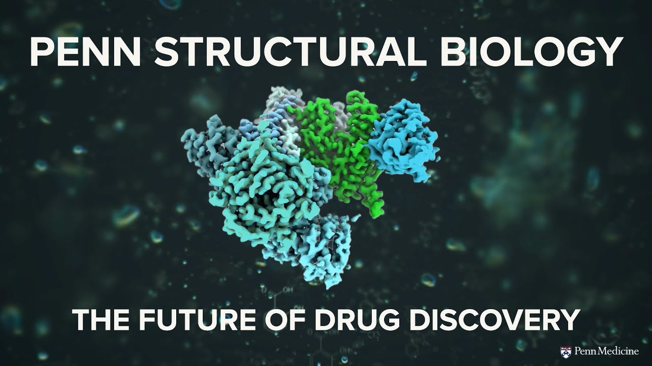
This ability to see individual components of the cell—proteins, nucleic acids such as RNA and DNA, lipids, and organelles as they go about their everyday work—is rapidly transforming the study of the biology behind health and disease.
Researchers at Penn Medicine are at the forefront of bringing this revolutionary imaging technique, known as cryogenic electron microscopy, or cryoEM, to improving patient health and quality of life by investigating causes and treatments for diseases such as Alzheimer’s disease, cancer, and influenza.
“With this technique, we are seeing a shift towards a detailed understanding of proteins and other components inside the cell, and this is enabling new questions to be answered,” said Vera Moiseenkova-Bell, PhD, a professor in the department of Systems Pharmacology and Translational Therapeutics and director of Penn Medicine’s Institute of Structural Biology (ISB).
With both cryoEM and other structural biology methods, the relatively new ISB, established in 2023 to study the shapes and functions of complex biological molecules, is beginning to realize how these insights can benefit clinical research, Moiseenkova-Bell said. “With these advances, I believe we're entering a new era of cellular structural biology.”
The ISB overall is home to 30 research laboratories that also study molecules using longer-established techniques like X-ray crystallography, NMR spectroscopy, single-molecule imaging, and computational biology. And the institute’s researchers are highly collaborative across other departments and centers at Penn, helping to bridge their discoveries to answer questions that can impact human health and disease and contribute to outcomes that matter for patients.
The particularly sharp ability to see biological molecules in action through cryoEM technology is filling in gaps in our knowledge of how their structures give rise to functions, said E. Michael Ostap, PhD, senior vice dean and chief scientific officer at Penn Medicine, whose research as a professor of Physiology explores the proteins that power cardiac muscle contraction.
“CryoEM gives a view inside cells that is like watching an entire football game, where other methods might show the kickoff and touchdown or the start and finish. CryoEM freezes thousands of players mid-action, each in a slightly different pose, and from these snapshots, scientists piece together the passes, tackles, and plays that make the entire game make sense.”
Ostap said that looking at molecular complexes with cryoEM is like watching an entire football game, where other imaging methods might show the kickoff and touchdown or the start and finish. CryoEM freezes thousands of players mid-action, each in a slightly different pose, and from these snapshots, scientists piece together the passes, tackles, and plays that make the entire game make sense.
“If you can actually see the different steps during an enzymatic reaction or other dynamic process,” Ostap said, “you can learn what components are important, as well as which components you might target in order to assist or inhibit important cell processes.”
Into the deep freeze
As the name suggests, cryoEM involves plunging proteins and other biological samples into cryogenic—that is, extremely cold—temperatures. The first step is to submerge the protein, nucleic acid, or molecular complex into a frosty concoction of liquid ethane that cools the specimen to about -188 degrees Celsius.
This rapid cooling turns the surrounding water into a glass-like solid rather than crystals of ice, which can damage the sample. The process traps the protein in a frozen cocoon that protects it from the electron beam, which normally would obliterate delicate biological structures.
Then the microscope fires a beam of electrons toward the protein. The electrons bounce off the protein’s atoms and fall onto a detector, where they create a shadow-like pattern. A computer turns these 2D snapshots into a 3D image. By averaging the patterns from thousands of identical proteins, the software can refine the image to deliver an atom-by-atom rendering of individual molecules, a process that researchers call “single particle analysis.”
First invented in the 1980s, cryoEM underwent vast improvements in image resolution in the early 2010s, leading to its adoption by major research centers such as Penn. Three early pioneers in this field garnered the 2017 Nobel Prize in Chemistry.
A related technique, cryo-electron tomography (cryoET), captures images of a single particle from multiple angles. The software then works out how to combine the images to reconstruct the 3D structure.
Compared to most other tools in the biologist’s toolkit, cryoET provides one big advantage: It allows researchers to see how proteins and other molecules do their work in more natural environments, including inside cells.
Preparing the sample is also easier than with the technique that was formerly the gold standard of molecular imaging, X-ray crystallography, known for its role in determining the structure of DNA in the 1950s. This method requires researchers to coax molecules into rigid crystals that may not accurately mimic their behavior in real life.
The combination of single particle analysis and cryoET with other techniques for exploring the machinery of human life—such as X-ray crystallography, nuclear magnetic resonance, mass spectrometry, artificial intelligence, and others—is especially powerful for creating a detailed understanding of human life and disease.
Penn’s two cryo-electron microscopes serve scientists across campus, in both biomedical research and the physical sciences and engineering. Housed at the university’s Beckman Center for Cryo-Electron Microscopy in the Krishna P. Singh Center for Nanotechnology, the facilities are used by up to 50 researchers across Penn. The equipment was acquired with support from the Arnold & Mabel Beckman Foundation and the National Institutes of Health.
To foster collaborations, Moiseenkova-Bell established seed grants that allow researchers from across the university to collaborate with Penn Medicine ISB researchers on projects involving cryoEM and cryoET.
Eventually, Moiseenkova-Bell hopes, structural biologists can combine tools to create a comprehensive understanding of the landscape of the cell, a sort of Google map of where every protein or major molecular complex can be identified. With such a map, researchers could ask what each component of the cell does, and how it contributes to keeping the body healthy.
Today, a flurry of major research findings at Penn Medicine in just the last few years are already showing how the institution’s investments are paying off: CryoEM is uncovering new understandings the mechanisms of diseases, smarter drug discovery, and fundamental insights into biological processes.
The study of diseases with cryoEM and cryoET includes revealing how parasites hijack our cells, how neurobiological disorders like Alzheimer’s harm the brain, and how the seemingly harmless “garbage trucks of the cell,” the lysosomes, play a role in disease.
New approaches to drug development are informing strategies for improving vaccines against flu, finding targets for treating cardiovascular disease, and blocking the body’s fat-producing enzymes that fuel the spread of cancer.
Over the long term, cryoEM discoveries about biological mechanisms, such as how chromosomes divide effortlessly during cell division, or how the protein actin lengthens or shortens during cancer metastasis, provide the ground for future therapeutics that promote health.
Explore the super-cool science of cryoEM
Seeing the mechanisms of disease with cryoEM
Understanding how a disease starts and gets going is essential to finding treatments—and imaging with cryoEM and cryoET is leading to such insights.
Zooming into drug discovery with cryo-microscopic science
Penn is at the forefront of using close-up imaging techniques to suggest new ways to match drugs to biological receptors like a key with a lock.
Biological insights of the future come from cryoEM today
Structural biologists are now more clearly seeing fundamental mechanisms of how cells function in the human body and across many forms of life.
Related articles
New frontiers in nipping cancer in the bud
The science of cancer interception is advancing at Penn Medicine with a new platform to develop cancer vaccines piling scientific strength upon strength.
Preserving Pennsylvania Hospital’s history ‘never gets old’
Stacey Peeples oversees the archives of the nation’s first chartered hospital, home to a nearly three-hundred-year collection of medical artifacts.