
Zooming into drug discovery with cryo-microscopic science
Close-up imaging techniques are suggesting new ways to match drugs to biological receptors with the precision of a key and lock.
At some level, getting the flu is a bit like being invaded by tiny aliens. Once the influenza virus infiltrates your cells, it rapidly replicates its genomic RNA and transcribes it into messenger RNA (mRNA), which provides the recipe to build viral proteins that are assembled into new viruses.
Newer methods of looking at the shapes of molecules are providing sharp new images of how these steps unfold during infection, suggesting new avenues for drug discovery for a variety of diseases. Researchers at Penn Medicine are at the forefront of bringing revolutionary imaging techniques, known as cryogenic electron microscopy (cryoEM), and cryo-electron tomography (cryoET), to understanding better ways to match drugs to the ways they can act on the molecules of life.
In the case of flu, which causes up to 650,000 deaths annually worldwide, researchers hope to apply this deeper understanding to building a universal flu vaccine or antiviral drug. This treatment could be used year after year, because it would target viral replication, an aspect of the virus’s function that evolves less frequently than the surface proteins targeted by today’s therapies.
Viral replication happens inside a molecular machine called the ribonucleoprotein complex. This multi-protein structure includes an RNA-copying enzyme called polymerase that makes millions of copies of the viral RNA and proceeds through the transcription process to produce viral proteins, all with the goal of packaging up a new alien army of infectious agents to unleash on the body.
But capturing high-resolution images of the ribonucleoprotein complex has been difficult, because some parts of the structure are highly flexible and can move around. The software that conducts single particle analysis struggles to average together images from flexible structures, because the software is looking for features that are similar across all the copies of the molecule.
Consider this example: If the software were averaging human faces, it would easily locate the mouth, nose, and eyes because these are always roughly in the same location. But what if the software were combining images of the entire human body, where each person could be doing something different, like waving a hand, or kicking up a leg? The software would average the body’s trunk but would struggle to produce crisp images of the limbs.
To meet this challenge, Ruchao Peng, PhD, a postdoctoral scholar working with Yi-Wei Chang, PhD, an assistant professor of Biochemistry and Biophysics and associate director of the Institute of Structural Biology, came up with two workarounds. First, the team created lab-made ribonucleoprotein complexes that have the improved rigidity needed for imaging in cryoEM.
This helped, but some parts of the structure are dynamic, especially during the active working process to produce RNA, so for their second workaround, they turned to cryoET to obtain images of the flexible parts from each complex.
By imaging thousands of flu ribonucleoprotein complexes using cryoEM and cryoET, the researchers could combine the snapshots into a movie to show how the entire machine worked.
What they saw astounded them. They saw in unprecedented detail the right-handed, double-stranded helix that made up the complexes. The enhanced resolution allowed them to, for the first time, see the viral genome RNA sitting in the minor groove of the helix, while the polymerase traveled down the helix like a train on its tracks to transcribe the viral RNA into mRNA.
They noted that throughout the entire transcription process, the viral RNA strand stayed protected inside the helix, with only a small fragment being progressively exposed to serve as the template for guiding mRNA transcription. This mechanism protects the viral RNA from degradation by host enzymes and maintains the structural integrity of the viral replication machinery, allowing repeated execution of the transcription process.
The images also revealed a potential Achilles heel in the process of viral replication that could lead to a new way to stop the flu virus from copying itself: There is a binding pocket between subunits along the helix where a small molecule could be inserted, like a key into a lock, to block the polymerase from doing its job. The team published the study in Science in May 2025. They are now screening small molecule compounds and have identified multiple candidates that can block this process, providing a new direction in the fight against flu.
A closer look: Figuring out flu’s Achilles heel
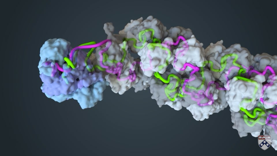
Penn Medicine researchers studied the first step in how the influenza virus converts its genetic instructions, in the form of RNA, into proteins that help it infect host cells. The transcription of viral RNA into messenger RNA takes place inside a molecular complex called a ribonucleoprotein (grey and white). Within this helical complex, an RNA-copying enzyme called a polymerase moves along the viral RNA, transcribing it into mRNA (long purple strand), which will later be translated into viral proteins. Source: Yi-Wei Chang laboratory, Penn Medicine.
Finding the best key for the lock
The search for small molecules that fit a binding site is a common approach for discovering drugs that interfere with disease.
Researchers at Penn Medicine are using cryoEM and cryoET to improve the odds of finding drug candidates.
Vera Moiseenkova-Bell and her team study membrane-bound ion channels, a hot target in the search for small molecules that can mediate a wide range of diseases.
Ion channels are proteins on the surfaces of cells that act like the city gates, permitting or denying charged atoms, or ions, from entering the cell. Once inside, these ions turn on cascades that regulate cellular processes.
A given channel can have roles in many functions and diseases. For example, one known as transient receptor potential vanilloid 2 (TRPV2) has possible roles in cell migration, cancer metastasis, immune cell regulation, neurodevelopment, and cardiovascular function.
Julia Rocereta, a graduate student working with Moiseenkova-Bell, used cryoEM to view TRPV2 when it was bound to a drug called probenecid, which is used as a medication to increase the body’s excretion of uric acid.
Visualizing the channel at near-atomic resolution, the researchers determined the location of drug binding. The images from cryoEM provide researchers with information they can use to optimize the shapes of other small molecule drugs that can fit in the binding pocket. The study was published in February 2025 in Nature Structural and Molecular Biology.
Rocereta said it was fulfilling to be among the first to explain how this drug is actually targeting this ion channel.
“We are doing drug discovery, but on a completely different level, starting from a structural perspective,” Rocereta said.
Other Penn Medicine researchers are looking at molecular structures and processes with an eye toward disrupting cancer. In the lab of Ronen Marmorstein, PhD, the George W. Raiziss Professor in the Department of Biochemistry and Biophysics, for example, Kollin Schultz, PhD, now a postdoctoral researcher, used cryoEM to look at the workings of an enzyme complex that churns out fatty acids, which are essential for growth and development, but also can be coopted by tumors to build the membranes of new cancer cells.
Schultz captured snapshots of the fatty acid synthase complex as it performed its task.
“We were finally able to get a more realistic picture of how it was working,” Schultz said, “and we are now able to make sense of a lot of the many years of biochemical data that hadn't quite added up into a single mechanism.”
Knowledge of this pathway could help researchers develop inhibitors of this complex, thus shutting down its contribution to cancer and fatty liver disease. The study, a collaboration with Kathryn E. Wellen, PhD, a professor of Cancer Biology at the Abramson Family Cancer Research Institute, was published in Nature in February 2025.
Marmorstein credits Penn with establishing an environment that encourages connections across disciplines, including structural biology, with numerous other areas of science and medicine.
“I think what's special about Penn is that it's really highly collaborative,” Marmorstein said. “People like to work together. This makes doing science a lot more fun, and it allows your work to go beyond where it would normally be able to go at an institution where the interactions were more limited.”
Explore the super-cool science of cryoEM
Freeze, image, cure
Researchers are capturing images of the biology inside our cells using cryogenic electron microscopy to inform how we understand and treat disease.
Seeing the mechanisms of disease with cryoEM
Understanding how a disease starts and gets going is essential to finding treatments—and imaging with cryoEM and cryoET is leading to such insights.
Biological insights of the future come from cryoEM today
Structural biologists are now more clearly seeing fundamental mechanisms of how cells function in the human body and across many forms of life.
Related articles
Preserving Pennsylvania Hospital’s history ‘never gets old’
Stacey Peeples oversees the archives of the nation’s first chartered hospital, home to a nearly three-hundred-year collection of medical artifacts.
Seeing the mechanisms of disease with cryoEM
Understanding how a disease starts and gets going is essential to finding treatments—and imaging with cryoEM and cryoET is leading to such insights.