
X-ray plates from 1896 give a snapshot of Penn’s place in history
A gift from the family of Penn physicist Arthur Goodspeed represents the beginning of a revolution in medicine that began at Penn.
For decades, the pair of glass X-ray plates from 1896 sat snugly wrapped in tissue paper, inside a Wanamaker department store box, in a spare closet in New Zealand.
“Arthur’s plates ... 2 early X-ray pictures,” reads the handwriting on the box.
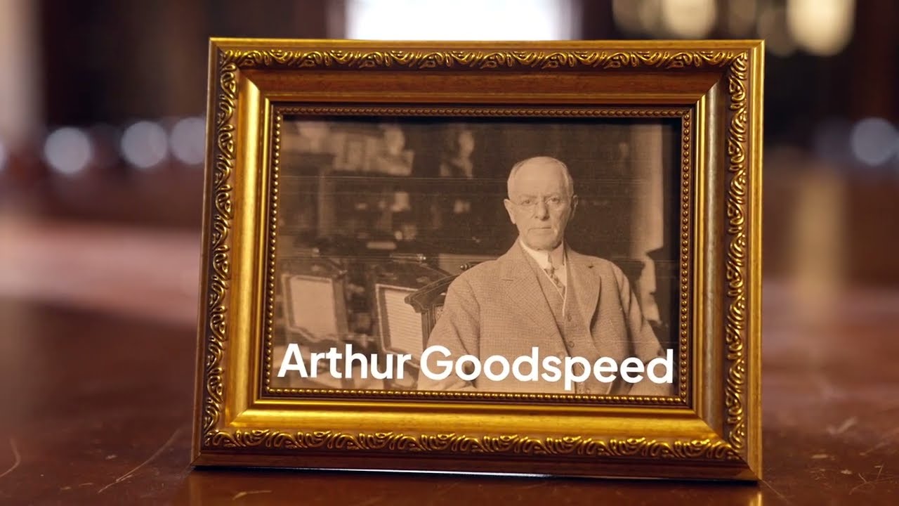
Arthur was Penn physics professor Arthur Goodspeed, who very well may have created the world’s first X-ray image at the University of Pennsylvania in 1890—entirely by accident. And those plates represent some of the world's earliest experiments with this type of imaging, shortly after X-rays were actually discovered in Germany in 1895.
As the story goes, in 1895, German physicist Wilhelm Röntgen published his discovery of a new type of radiation that could pass through solid objects and create images that could be printed as negatives onto photographic plates. He called them X-rays to signify the unknown nature of the radiation; they would also be called “Röntgen rays.”
It was at that point that Goodspeed remembered a mysterious incident. Nearly six years earlier, he and Philadelphia photographer William Jennings had been trying to photograph electrostatic discharges from an induction machine onto glass photographic plates—then used instead of film—without a camera. Then Goodspeed got out Penn’s collection of Crookes tubes—vacuum-sealed containers used to study cathode rays—to show his photographer friend.
Unbeknownst to them, the tube was emitting radiation near a glass photographic plate on which two coins had been laid. When Jennings developed the plates, he reported two “mysterious” discs that couldn’t be explained.
Goodspeed forgot about it until Röntgen’s discovery, when he realized how that mysterious image had been made.
In January 1896, Goodspeed recreated Röntgen’s experiments using a coin purse and two coins, outlining his attempts in the March 13, 1896, issue of Science. There, he also recounted the incident from six years earlier.
Goodspeed wrote that he and Jennings “wish to claim no credit for the interesting accident, but the fact remains that without a doubt the first Röntgen picture was produced on February 22, 1890, in the physical lecture room of the University of Pennsylvania.”
Almost immediately after those coin experiments, Goodspeed teamed with Penn Medicine surgeons to produce some of the first X-rays of patients. It was the birth of diagnostic imagery at Penn.
“It was very quick that they envisioned that medicine was going to be a good application for the X-ray,” said radiologist Mitchell Schnall, MD, PhD, Penn Medicine’s senior vice president for data and technology solutions. “Very quickly, Penn became a large center for radiology and X-ray.”
A tangible artifact from 1896
Through the decades, two X-ray plates from Goodspeed’s early experiments were kept by various family members, most recently at the home of granddaughter Dianne Goodspeed Halliday in New Zealand. Measuring about 3 by 4 inches, each plate shows the outline of a coin purse with two coins inside. They are estimated to be from January or February 1896 based on the Science article and when more advanced images were made.
In recent years, Halliday and other family members decided Penn should have their pieces of history.
“We all felt that the plates were of enough historical interest to be preserved,” Halliday said.
A cousin in Swarthmore—Goodspeed’s great-granddaughter, Sharon Goodspeed, who loves telling people about her great-grandfather’s place in medical history—agreed.
“They're of great significance, so they should be kept well and stored well and shared so that people can learn more about my great-grandfather.”
The donation nearly went awry during Halliday’s trip to the states, when her backpack containing the plates was stolen from a hotel coffee shop in San Francisco. The thief ran off with an iPad but left the plates unharmed, except for a small chip.
Halliday and Goodspeed then delivered them to Penn, still in the Wanamaker box, along with some other family items related to Penn’s history.
Penn University Archivist John Bence was thrilled.
“In my experience as an archivist, singular objects that are evidence of a scientific discovery are really rare,” Bence said. “Scientists move on pretty quickly—always iterating and improving—and that retrospective view is rarely taken. So to have these is really exciting.”
At the forefront of imaging in medicine for 128 years and counting
X-rays were a revolution for medical diagnostics, with Goodspeed and Penn playing a central role. Penn’s Radiology division, which opened in 1897, was believed to be the first in the U.S.
At that time, it was easy for Goodspeed and others to experiment and to quickly translate discoveries for clinical practice, Schnall said. And although the environment has drastically changed, the spirit of innovation has driven the radiology department to this day.
As an example, Schnall points to his own experience as a young faculty member in the 1990s, when he and his colleagues built the technology to do more advanced breast magnetic resonance imaging (MRI) studies than what the standard equipment allowed.
“In those days, if you came to Penn for an MR exam, chances are, the coil that was used in that exam was built by us, and chances are, the pulse sequence that was run to collect the images was written by us on top of what the manufacturer provided, so it was an exciting time—probably much like these guys,” he said.
And the technology for those MRI scans—arguably the most sophisticated imaging modalities radiologists have to this day—was built on the fundamentals of X-ray and radiology imaging laid by Goodspeed, Schnall said.
Connecting radiology’s history to the future
In September 2025, the Goodspeed plates and the story behind them were shared with Perelman School of Medicine leaders, at a meeting where the group also welcomed Penn’s newest Radiology chair, Pari Pandharipande, MD, MPH—connecting the department’s past with its present and future.
Pandharipande expressed profound gratitude to the Goodspeed family for appreciating the significance of the X-ray plates and wanting Penn to have them. She noted the plates exemplify a culture she is proud to join.
“This department in particular has really moved the field of radiology forward in modality after modality,” she said. “The fact that Goodspeed did this here, before 1900—it resonates with that culture of innovation in the department. That's really one of the things that has made me very excited to come to Penn.”
Upon hearing the story, Abramson Cancer Center Director Robert Vonderheide, MD, DPhil, immediately looked up the Science article from 1896 where Goodspeed described his experiments to replicate Röntgen’s discovery. Vonderheide was struck by the thread connecting more than a century of science and medicine at Penn.
“I'm a faculty member at the University of Pennsylvania. So was he. We all want to publish in that journal,” Vonderheide said. “It's just remarkable how the spirit of discovery hasn't really changed. We share that as the descendants of this tradition.”
Related articles
The tinkerer-turned-tech leader
Mitchell Schnall, MD, PhD, is using his insights from technology in radiology to solve problems and scale up changes in the health system.
Replicant’s dream to reality: Imaging AI at Penn Medicine
A new Penn Medicine-developed AI tool is providing a new level of diagnostic precision to physicians reviewing patient scans.